ADJUSTABLE SOURCE TO IMAGE DISTANCE HELPS IMPROVE RADIOGRAPHIC INTERPRETATION
MyVet i72 Veterinary x-ray table system
Veterinarians benefit in clinical interpretations with variable SID / 72” horizontal beam imaging digital X-ray system

MyVet i72 variable 40” – 72” SID Veterinary X-ray table system designed to improve diagnostic image interpretation by reducing geometric image distortion effects typically associated with fixed 40” SID systems. Horizontal beam imaging capability facilitates weight bearing exams as well as imaging air/fluid level radiographs of the chest or abdomen when clinically indicated.
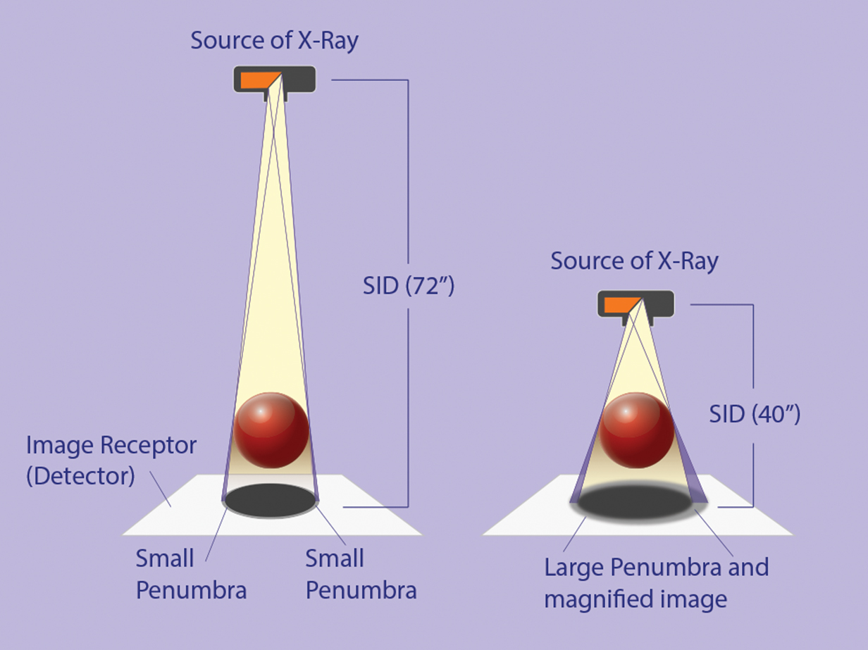
Overall radiographic image formation is primarily a function of two geometric factors: magnification and penumbra. The i72 variable 40” – 72” SID in both vertical as well as horizontal planes overcomes both.
 While magnification does not negatively impact radiographic quality, if absolute measurement of size is important or a greater field of view is required to capture more of an anatomical structure then a fixed 40” SID can provide, the i72 variable SID facilitates the capture of a complete extremity, for example, from shoulder-to-paw or hip-to-paw or by minimizing x-ray beam divergence that causes magnification.
Additionally, the i72 variable SID reduces the effect of penumbra on image sharpness. As SID increases, the blurring effects of penumbra on image edge sharpness is reduced as a result of a more parallel x-ray beam imaging fine anatomic structures such as lung vascularity, bone trabeculae or, in particular, bone lesion margins. Radiographically, the sharper the lesion margin between normal and abnormal bone, the more likely the lesion is benign.
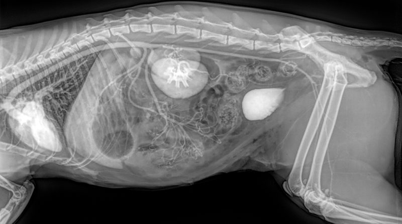
During emergent care for animals in respiratory distress, obtaining radiographs is important but recumbent views only exacerbate the problem as the animal labors to breathe. If images are immediately required for a more concise clinical diagnosis, the i72 horizontal beam capability helps secure right and left lateral images in the upright position thereby reducing further breathing difficulty precipitated by recumbent positioning.
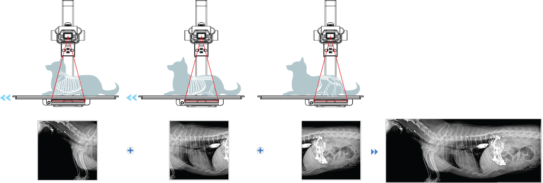
System functionality includes image stitching at 40” or 72” SID with auto table positioning. Dynamic table positioning and sync positioning allows the X-ray tube head to maintain a 40” SID with the table as it is raised or lowered. Quiet motor controls and table Bucky movement reduces animal startle reflexes that help vet technicians quickly and efficiently complete an imaging exam.
“The i72 digital radiographic table is the first x-ray system that actively allows Veterinarians to use X-ray beam geometry principles to their advantage”, said Bill Nicholas, Director of Marketing and Corporate Communications. “Instead of being bound by these principles, they may use them to improve their ability to make better diagnoses”, he finished